ClariPulmo
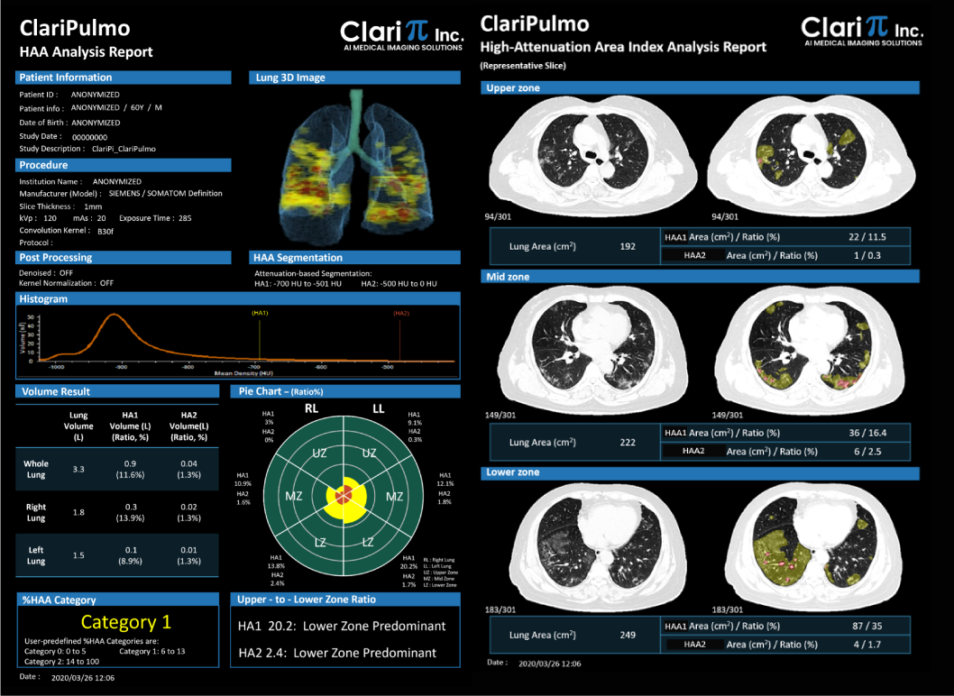
ClariPulmo is a non-invasive image analysis software for use with CT images which is intended to support the quantification of lung CT images. The software is designed to support the physician in the diagnosis and documentation of pulmonary tissue images (e.g., abnormalities) from the CT thoracic datasets. The software provides automated segmentation of the lungs and quantification of low-attenuation and high-attenuation areas within the segmented lungs by user predefined Hounsfield unit thresholds. The software displays by color the segmented lungs and analysis results. ClariPulmo provides optional denoising and kernel normalization functions for improved quantification of lung CT images in cases when CT images were taken at low-dose conditions or with sharp reconstruction kernels.

ClariPulmo Benefits
ClariPulmo Provides
- Lungs are automatically segmented using a pre-trained deep learning model. Report includes results with location as well as 3D rendered image
- Optional Kernel Normalization function provides an image-to-image translation from a sharp kernel image to a smooth kernel image for improved quantification of lung CT images
- Optional denoising function provides an image-to-image translation from a noisy low dose image to a noise-reduced enhanced quality image for improved quantification of lung images
- ClariPulmo provides summary reports for measurement results that contain color overlay images for lung tissues as well as table and charts displaying analysis results
LAA & HAA Analysis
- LAA Analysis provides quantitative measurement of a pulmonary tissue image with low attenuation areas (LAA). This feature supports the physician in quantifying lung tissue image with low attenuation area
- HAA Analysis provides quantitative measurement of pulmonary tissue image with high attenuation areas (HAA). This feature supports the physician in quantifying lung tissue image with high attenuation area
Operating Costs Reduction
- Integrate with existing CT scanners meeting specifications
- Double solution package at one unit price
How It Works
Fully compliant with DICOM standards, ClariPulmo allows easy and seamless integration with CT scanners and PACS systems meeting specifications.
We are looking for Hospitals/Clinics, Radiologists, PACS Companies, Manufacturers, Dealers for New or Used Medical Equipment


